We recently received an inquiry about the feasibility of modeling biological structures such as the cochlea of the human ear with the AMaze programs. The assumption is that slice data (such as an MRI scan) is available to define the morphology of the structure. It turns out that such modeling would be straightforward with MetaMesh, which already includes features to convert three-dimensional image information into a conformal hexahedron mesh. In this article, I will discuss how the conversion could be carried out and some of the limitations of the process.
Think of a medical image as a series of planar images in x-y at a number Nz of positions in z. A single plane consists of gray-scale values (0-255) in voxels over an array Nx by Ny. The crux of the problem is whether the brightness values can be related to physical properties of the medium. For an electric-field solution the properties would be relative dielectric constant and/or conductivity. There are three levels of conversion solutions:
- The cochlea model would be relatively easy. The goal is to identify the boundaries of the void inside the organ which would have good contrast with the surrounding tissue. The conversion could be done automatically by a interpreter script, assigning a value of 0.0 to the void and 1.0 to tissue.
- A model of a section of the spine would be of moderate difficulty. There is good contrast between the bone, cartilage, nerves and muscles. The model would probably require a combination of automatic analysis and human corrections.
- A model of the brain would be difficult and time-consuming. There is considerable structure, and the gray-scale values overlap in different structures. It would probably be necessary for a knowledgeable human to outline critical regions in each layer.
To illustrate the process, I constructed a mathematical model of a complex duct. The duct is a cylinder with a harmonic modulation of radius along z (like a vacuum bellows). At the same time, it twists in a helical pattern. I used the following equations for the boundary of the duct:
Helical displacement of the centerline at position z:
xc = D*cos(TwoPi*z/Zr)
yc = D*sin(TwoPi*z/Zr)
Radius of the duct at z:
R = R0 + Dr*sin(TwoPi*z/Zh)
Displacement from the center at position (x,y) at z
rd = sqrt((x-xc)*(x-xc)+(y-yc)*(y-yc))
A voxel with center-of-mass at (x,y) is assigned the value of tissue (1.0) if rd > R and void (0.0) if rd < R. I wrote a Perl script to create a MetaMesh image file with parameters
XMin = -5.0, XMax = 5.0, Nx = 100
YMin = -5.0, YMax = 5.0, Ny = 100
ZMin = -10.0, ZMax = 10.0, Nz = 200
Here is the full MetaMesh script to read the image file and create the shaped void (region 2) inside the tissue (Region 1). The format of the Type Image command is covered in detail in Chap. 20 of the MetaMesh manual.
* File ORGAN.MIN
Global
  XMesh
    -5.00  5.00   0.10
  End
  YMesh
    -5.00  5.00   0.10
  End
  ZMesh
    -10.00  10.00  0.10
  End
  Smooth 0
End

Part
  Type Box
  Region 1
  Fab  10.00  10.00  20.00
End
Part
  Type Image ORGAN
    0.00   0.10   2
  End
  Fab -4
End
EndFile
Figure 1 shows the mesh created for the duct after 4 cycles of surface smoothing for R0 = 3.0, D = 0.75, Dr = 0.25, Zr = 10.0 and Zh = 3.0. The left side shows the division in one z-plane, while the right-hand side shows the three-dimensional shape of the duct wall.
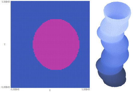
Figure 1. Mathematical simulation of a biological structure.
LINKS
